The human eye is a remarkable organ that allows us to experience the world in vivid detail. Behind the visible part of the eye lies the retina, a delicate layer of tissue that plays a critical role in vision. While most people rarely think about their retina, it is essential to maintain its health to prevent serious eye conditions. One such condition that can affect the retina is lattice retinal degeneration.
Lattice Retinal Degeneration is a condition that affects the peripheral retina, the outer edges of the retina that are not directly responsible for sharp central vision but are crucial for overall eye health. This condition often develops slowly and may not cause noticeable symptoms initially. However, if left undetected, it can increase the risk of retinal tears or detachment, which can lead to vision loss.
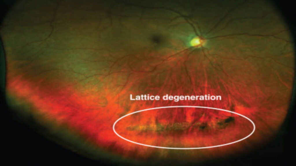
What is Lattice Retinal Degeneration?
Lattice retinal degeneration is characterized by thinning and weakening of the retinal tissue in specific areas, forming a lattice-like pattern. These lattice-shaped areas appear as elongated, crisscrossing lines or bands in the peripheral retina. The condition is usually diagnosed during a routine eye exam using specialized equipment that allows an ophthalmologist to carefully examine the retina.
While lattice retinal degeneration can occur in anyone, certain factors increase the likelihood of developing it. People who are highly nearsighted (myopic), those with a family history of retinal problems, and older adults are more prone to this condition. It is also more common in both eyes rather than just one. Despite the changes in retinal tissue, many individuals may not experience any vision problems for years, which is why regular eye examinations are crucial.
Causes and Risk Factors
The exact cause of lattice retinal degeneration is not fully understood, but several factors contribute to its development:
-
Genetics: A family history of retinal degeneration or retinal detachment can increase susceptibility.
-
High Myopia: Individuals with severe nearsightedness are at higher risk because their eyes are elongated, stretching the retina and making it more fragile.
-
Age: Lattice degeneration is more commonly observed in middle-aged and older adults.
-
Trauma: Past eye injuries or surgeries can contribute to the development of lattice degeneration.
-
Other Eye Conditions: Some retinal disorders and systemic diseases may increase vulnerability.
Understanding these risk factors is important because it helps patients and doctors identify who may benefit from more frequent retinal screenings.
Symptoms of Lattice Retinal Degeneration
One of the challenging aspects of lattice retinal degeneration is that it often remains asymptomatic until complications arise. Many people do not experience any warning signs. However, when symptoms do appear, they may include:
-
Flashes of light (photopsia): Brief flashes of light in the peripheral vision may indicate retinal irritation or the beginning of a tear.
-
Floaters: Small specks, lines, or shadows that drift across the field of vision may signal vitreous changes affecting the retina.
-
Peripheral vision loss: Gradual narrowing of the visual field can occur if retinal changes progress.
-
Sudden visual changes: A sudden shadow or curtain in vision is a medical emergency, possibly indicating retinal detachment.
Because early stages often produce no symptoms, proactive eye examinations are the most reliable method for detecting lattice retinal degeneration.
How is Lattice Retinal Degeneration Diagnosed?
Diagnosis begins with a comprehensive eye examination performed by an ophthalmologist or retina specialist. During the exam, the doctor uses specialized instruments to inspect the retina carefully. Common diagnostic techniques include:
-
Dilated Eye Exam: Eye drops are used to dilate the pupils, allowing the doctor to view the retina more clearly. This is the most common way to detect lattice degeneration.
-
Ophthalmoscopy: A handheld instrument called an ophthalmoscope allows direct examination of the retina.
-
Fundus Photography: High-resolution images of the retina can document areas of lattice degeneration and monitor changes over time.
-
Optical Coherence Tomography (OCT): OCT provides detailed cross-sectional images of the retina to evaluate its thickness and structure.
-
Ultrasound Imaging: If the retina is difficult to examine due to cataracts or other obstructions, ultrasound can help detect tears or detachments.
Early detection through these diagnostic tools is essential because it allows timely intervention, reducing the risk of complications.
Complications Associated with Lattice Retinal Degeneration
While lattice retinal degeneration itself may not immediately threaten vision, it can lead to serious complications if not monitored:
-
Retinal Tears: Weakened areas of the retina are prone to small breaks or tears. These tears can allow fluid to seep under the retina, potentially leading to detachment.
-
Retinal Detachment: A retinal detachment occurs when the retina separates from the underlying tissue, causing loss of vision. This condition is a medical emergency that requires prompt treatment.
-
Vitreous Hemorrhage: In some cases, tears in the retina can lead to bleeding into the vitreous gel, causing floaters or blurred vision.
Regular follow-up with an eye specialist is crucial for patients with lattice retinal degeneration to monitor for these complications.
Treatment Options
Treatment for lattice retinal degeneration depends on the severity and associated risks. Not every patient requires intervention, especially if the degeneration is stable and there are no tears. However, in cases where the retina is at high risk of tearing or detachment, preventive measures may be recommended.
-
Observation: Most cases are carefully monitored through regular eye exams. Patients are advised to report any new symptoms immediately.
-
Laser Photocoagulation: This procedure uses a focused laser to create small burns around retinal tears or weak areas. The burns form scar tissue that seals the retina and reduces the risk of detachment.
-
Cryotherapy: In some cases, freezing therapy is used to secure the retina, especially when laser treatment is not suitable.
-
Surgery: If a retinal tear or detachment occurs, surgery may be necessary. Procedures such as scleral buckling, vitrectomy, or pneumatic retinopexy are performed to repair and reattach the retina.
Preventive treatment is often highly effective in reducing the risk of serious vision loss, which is why early detection is so important.
Lifestyle and Home Care Tips
While medical treatment is essential for lattice retinal degeneration, lifestyle adjustments can also support eye health and reduce risks:
-
Regular Eye Exams: Individuals at risk should have comprehensive retinal exams at least once a year.
-
Protect Eyes from Trauma: Wearing protective eyewear during sports or risky activities can prevent injuries that may worsen retinal conditions.
-
Manage Systemic Health: Conditions such as diabetes and hypertension can affect retinal health, so controlling these diseases is important.
-
Know the Warning Signs: Immediate medical attention is necessary if you notice sudden flashes, floaters, or visual shadows.
Although lifestyle changes cannot reverse lattice degeneration, they help protect overall eye health and support early detection of complications.
Who Should Get Screened?
Screening for lattice retinal degeneration is particularly important for individuals with the following risk factors:
-
High myopia (severe nearsightedness)
-
Family history of retinal tears or detachment
-
Previous eye trauma or surgery
-
Age above 40
-
History of retinal disorders
Early screening allows ophthalmologists to identify weak areas in the retina and implement preventive measures before serious problems occur.
The Role of Technology in Detection
Advances in eye care technology have made detecting lattice retinal degeneration more precise and less invasive. Modern imaging techniques, such as optical coherence tomography and fundus photography, allow doctors to monitor subtle changes over time. These tools can identify areas of thinning that might not be visible with a standard eye exam alone.
Telemedicine and AI-assisted retinal imaging are also becoming increasingly useful. These technologies can analyze retinal images and flag potential issues, making early detection accessible to a broader population.
Prognosis and Long-Term Outlook
For many individuals, lattice retinal degeneration can remain stable for years without causing major vision problems. The key to maintaining vision is early detection and monitoring. With timely intervention, the risk of retinal detachment can be minimized, and patients can continue to enjoy good vision throughout life.
Even if preventive procedures like laser photocoagulation are necessary, most patients retain excellent vision after treatment. Regular check-ups and awareness of symptoms are crucial components of long-term care.
Conclusion
Lattice retinal degeneration is a condition that may not cause noticeable symptoms in its early stages but carries the risk of serious complications such as retinal tears and detachment. Understanding the risk factors, getting regular eye exams, and seeking timely medical attention are critical for preserving vision. Advances in imaging and treatment options make it possible to manage this condition effectively, often preventing significant vision loss.
If you are concerned about lattice retinal degeneration or have risk factors for retinal complications, consulting a specialist is essential. Dr. Mandeep Lamba, a renowned eye care professional, emphasizes the importance of early detection and regular monitoring. With proper care and guidance, patients can maintain healthy vision and reduce the risk of severe retinal problems.